Friday 7 August 2015

SAQ - Practice Paper 3

1. A 55 year old gentleman attends the ED with vertigo. He feels as though the room is spinning and he has intractable vomiting. When you enter the room he is lying still on the bed, every time he moves his head he feels like he is going to fall and vomits.


He has no previous medical illness and until this morning he was well except for a minor upper respiratory tract infection.


  1. What is the likely diagnosis?

  2. Give 8 other causes of vertigo.

  3. You examine the patient and identify no evidence of a central cerebellar lesion. What are 6 signs of cerebellar involvement?

  4. Name 2 drugs, with doses which you could use for this patient.

Answers:

Answers:


1. What is the likely diagnosis?
  • Viral Labyrinthitis

2. Give 8 other causes of vertigo.


  1. Meniere"s disease (vertigo, tinnitus and deafness)

  2. Benign positional vertigo (elderly, lasts approximately 2 mins with positional change)

  3. Otitis media

  4. Cholesteatoma

  5. Acoustic neuroma (giddiness more than vertigo, V, VI and VIII cranial nerve palsy, ipsilateral cerebellar signs, loss of corneal reflex)

  6. CVA

  7. Trauma

  8. Wax or foreign body in the ear

  9. MS

  10. Alcohol intoxication

  11. Ramsay Hunt syndrome

3. You examine the patient and identify no evidence of a central cerebellar lesion. What are 6 signs of cerebellar involvement?


The mnemonic DANISH will help to remember them:


  1. Dysdiadochokinesis/ dysarthria

  2. Ataxia (gait and posture)

  3. Nystagmus

  4. Intention tremor/ past pointing

  5. Slurred, staccato speech

  6. Hypotonia/hyperreflexia

4. Name 2 drugs, with doses which you could use for this patient.


  1. Prochlorperazine: 5 mg PO, 3 mg buccal or 12.5 mg IM

  2. Betahistine: 8 mg PO

  3. Cyclizine: 50 mg PO/IM/IV


2. According to ATLS


  1. List 6 immediate life threatening conditions in chest trauma

  2. Name 8 potential life threatening chest injuries which might be found on the secondary survey.

  3. Describe how you would perform pericardiocentesis.

  4. A 70 Kg patient has just been intubated. At what minute volume would you set the ventilator?

Answers:

Answers:


1. List 6 immediate life threatening conditions in chest trauma
  1. Tension pneumothorax

  2. Massive haemothorax

  3. Cardiac tamponade

  4. Open pneumothorax

  5. Flail chest

  6. Airway obstruction

2. Name 8 potential life threatening chest injuries which might be found on the secondary survey.


  1. Cardiac contusion

  2. Lung contusion

  3. Diaphragmatic injury/rupture

  4. Simple pneumothorax

  5. Oesophageal rupture

  6. Tracheo-bronchial disruption

  7. Traumatic aortic rupture

  8. Mediastinal traversing wound

3. Describe how you would perform pericardiocentesis.


  • Cardiac monitoring

  • 2 cm inferior to xiphisternum

  • Aim for left shoulder

  • Advance withdrawing syringe and withdraw needle if ST segment elevation on ECG


4. A 70 Kg patient has just been intubated. At what minute volume would you set the ventilator?


Minute volume = Tidal volume x respiratory rate ( 500 ml x 12) = 6L/min

Normal range - 5-8 L/min


More info: Patient.info (pericardiocentesis)


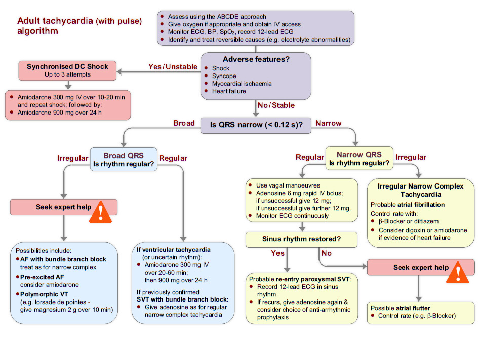
3. A 66 year old man brought to the ED . he has been experiencing palpitations. He is comfortable at rest. His heart rate is 160/min.


  1. What are 4 signs of instability?

  2. If a patient displays these signs, what is the immediate course of action? One drug and one non drug with doses as required?

  3. The ECG shows narrow complex tachycardia which is irregular. What key feature in the history will determine your management?

  4. Which drug can be used in treatment of atrial fibrillation according to the 2010 resuscitation council guidelines?

  5. On the bradycardia algorithm, what features indicate increased risk of asystole?

  6. Outline initial treatment of bradycardia which has failed to respond to an initial dose of atropine. Include doses.

Answers:

Answers:


1. What are 4 signs of instability?
  • Shock – hypotension (systolic blood pressure < 90 mmHg), pallor, sweating,

    cold, clammy extremities, confusion or impaired consciousness.

  • Syncope – transient loss of consciousness due to global reduction in blood

    flow to the brain.

  • Myocardial ischaemia – typical ischaemic chest pain and/or evidence of

    myocardial ischaemia on 12-lead ECG.

  • Heart failure – pulmonary oedema and/or raised jugular venous pressure

    (with or without peripheral oedema and liver enlargement).

2. If a patient displays these signs, what is the immediate course of action? One drug and one non drug with doses as required?


  1. Synchronised DC shock up to 3 attempts

  2. Then Amiodarone 300 mg IV over 10-20 minutes followed by 900mg over 24hrs.


ALS Book Says:
  • Carry out cardioversion under general anaesthesia or conscious sedation, administered by a healthcare professional competent in the technique being used. Ensure that the defibrillator is set to synchronised mode.

  • For a broad-complex tachycardia or atrial fibrillation, start with 120-150 J biphasic shock (200 J monophasic) and increase in increments if this fails.

  • Atrial flutter and regular narrow-complex tachycardia will often be terminated by lower energies: start with 70-120 J biphasic (100 J monophasic)".


3. The ECG shows narrow complex tachycardia which is irregular. What key feature in the history will determine your management?


  • Onset of arrhythmia <48 hrs. If yes, cardioversion. If no, rate control

4. Which drug can be used in treatment of atrial fibrillation according to the 2010 resuscitation council guidelines?


  • Beta Blocker ( Bisoprolol 2.5 to 5 mg PO, Metoprolol 5mg IV)

  • Diltiazem or Verapamil if beta blocker in contraindicated or not tolerated.

  • Digoxin 500 microgram loading dose ( if evidence of heart failure)

  • Amiodarone 300mg over 20-60mins followed by 900mg over 24 hrs.

5. On the bradycardia algorithm, what features indicate increased risk of asystole?


  • Recent asystole

  • Mobitz II AV block

  • Complete heart block with broad QRS

  • Ventricular pause > 3 s

6. Outline initial treatment of bradycardia which has failed to respond to an initial dose of atropine. Include doses.


  • Atropine 500mg IV to a maximum of 3mg

  • Isoprenaline 5 microgram/min

  • Adrenaline 2-10 microgram/min

  • Transcutaneous pacing

  • Alternate drugs: Dopamine, Aminophyline, Glucagon (beta blocker or Ca channel blocker toxicity), Glycopyrrolate
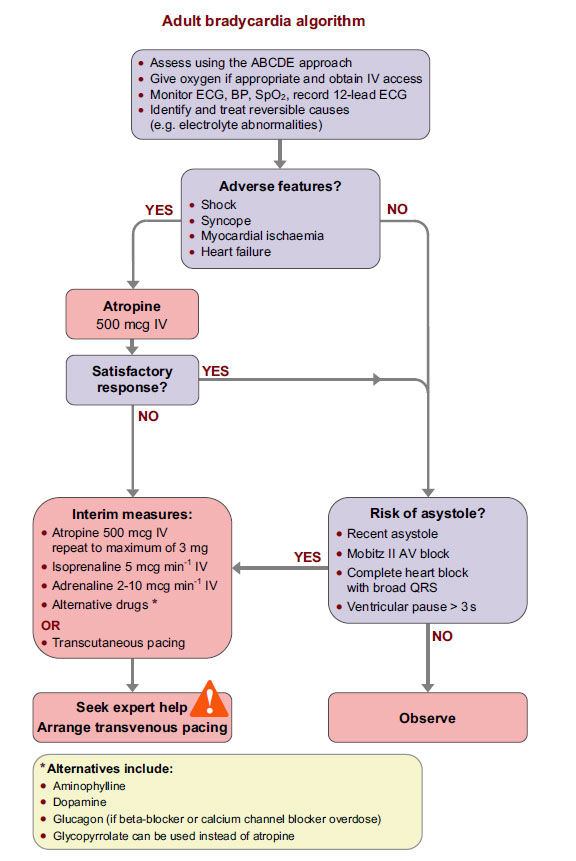

Resus Council Peri-arrest Arrhythmias Guidelines

4. A 35 year old builder attends the ED with a wound on his left thigh which he sustained on some barbed wire yesterday. He is diabetic, on insulin and has no known allergies. He is fully covered for tetanus vaccinations and noticed this morning that his wound, a 2cm superficial wound had become painful and red. The triage nurse marks the 1cm x 2cm area of redness and directs him back to the waiting room. You call him into the cubicle 2 hours later to find him looking unwell. He is pyrexial and looks toxic. The erythema has spread and is now blistered although he says he can"t feel is as much now. His pulse is 110/min and BP of 94/64.


You examine the leg and find it as you see it below.


  1. What is the likely diagnosis?

  2. Give 4 features in history and examination would help with the diagnosis.

  3. What are the usual causative organisms?

  4. Outline your management of this patient.

Answers:

Answers:


1. What is the likely diagnosis?
  • Necrotising fasciitis

2. Give 4 features in history and examination would help with the diagnosis.


  1. Rapidly spreading erythema

  2. Dusky purplish discolouration at wound

  3. Intense and severe pain which may seem out of proportion to any external signs of infection on the skin

  4. Anaesthesia to affected area at later stage due to nerve damage

  5. Putrid discharge/bullae/tissue necrosis

  6. Crepitation felt on tissue due to gas forming infection

  7. Clinical signs of shock/fever/toxic

3. What are the usual causative organisms?


  1. Group A haemolytic streptococcus (Streptococcus pyogenes)

  2. Staphylococcus aureus

  3. Clostridium perfringens

  4. Bacteroides fragilis

  5. Peptostreptococcus

  6. Colliforms

  7. Proteus

  8. Klebsiella

4. Outline your management of this patient.


  • Transfer the patient to resuscitation area

  • High flow oxygen with 15L rebreather mask

  • IV access and 1L of 0.9% Sodium Chloride stat

  • Blood for U&E, FBC, CRP, Culture & Sensitivity, ABG, Urinalysis

  • High doses of antibiotics: Benzylpenicillin plus clindamycin plus gentamicin

    If penicillin-allergic, meropenem plus clindamycin plus gentamicin

  • Urgent Plastics/Orthopaedic review


The Laboratory Risk Indicator for Necrotizing Fasciitis (LRINEC) score can be utilised to risk stratify people having signs of cellulitis to determine the likelihood of necrotising fasciitis being present.

It uses six serologic measures: C-reactive protein, total WBC count, haemoglobin, sodium, creatinine and glucose. A score greater than or equal to 6 indicates that necrotising fasciitis should be seriously considered.

The scoring criteria are as follows:


  • CRP (mg/L) ≥150: 4 points

  • WBC count (×10³/mm³)
    • <15: 0 points

    • 15–25: 1 point

    • >25: 2 points


  • Haemoglobin (g/dL) >13.5: 0 points 11–13.5: 1 point <11: 2 points

  • Glucose (mmol/L) >10: 1 point

  • Sodium (mmol/L) <135: 2 points

  • Creatinine (umol/L) >141: 2 points

  • Glucose (mmol/L) >10: 1 point

As per the derivation study of the LRINEC score, a score of ≥ 6 is a reasonable cut-off to rule in necrotising fasciitis, but a LRINEC < 6 does not completely rule out the diagnosis. Diagnoses of severe cellulitis or abscess should also be considered due to similar presentations.


Patient.infoPublic Health England


5. An 8 year old boy attends your emergency department having fallen out of a tree. His only injury is to his right ankle, which is swollen and painful. The X-ray is as shown below.


  1. Describe the X-ray appearance.

  2. What is the name of the classification of fracture of this type, and what type is this?

  3. Describe the other types of fracture in this classification.

  4. What will be the disposal of this patient and why?

Answers:

Answers:


1. Describe the X-ray appearance.
  • Fracture through the epiphysis and epiphyseal plate of the distal tibia.

2. What is the name of the classification of fracture of this type, and what type is this?


  • Salter Harris fracture, type 3

3. Describe the other types of fracture in this classification.


  • Type I - Slip at the growth plate

  • Tupe II - through the metaphysis and growth plate

  • Type III - Through epiphysis and growth plate

  • Type IV - through both epiphysis and metaphysis

  • Type V - crush at the growth plate

4. What will be the disposal of this patient and why?


  • Referral to orthopaedics on-call for fixation and there is articular involvement.

More info


6. A 60 year old man presents to your department having tripped over a dog. He has no other injuries except for the fracture of his right femur as shown


  1. Describe the X-ray.

  2. What is the diagnosis? What biochemical abnormality would your expect in the blood?

  3. How would you access the cognitive function of this gentleman.

Answers:

Answers:


1. Describe the X-ray
  • Transverse fracture of the femur

  • Complete transition

  • Proximal displacement of the distal fracture fragment

  • Bone shows patchy osteolytic and sclerotic areas

2. What is the diagnosis? What biochemical abnormality would your expect in the blood?


  • Paget"s disease

  • Increased alkaline phosphatase and rest are normal.

3. How would you access the cognitive function of this gentleman.


QuestionScore
What is your age? (1 point)
What is the time to the nearest hour? (1 point)
Give the patient an address, and ask him or her to repeat it at the end of the test. (1 point)e.g. 42 West Street
What is the year? (1 point)
What is the name of the hospital or number of the residence where the patient is situated? (1 point)
Can the patient recognize two persons (the doctor, nurse, home help, etc.)? (1 point)
What is your date of birth? (day and month sufficient) (1 point)
In what year did World War 1 begin? (1 point)(other dates can be used, with a preference for dates some time in the past.)
Name the present monarch/dictator/prime minister/president. (1 point)(Alternatively, the question "When did you come to [this country]? " has been suggested)
Count backwards from 20 down to 1. (1 point)

A score of 7-8 or less suggests cognitive impairment at the time of testing, although further and more formal tests are necessary to confirm a diagnosis of dementia, delirium or other causes of cognitive impairment.

More info on Paget"s Disease


7. A 75 year old man presents with 5 weeks history of back pain and gradual loss of mobility. His daughter says that whilst he used to be completely independent and walk several miles a day, he now needs help around the house and his exercise tolerance is reduced to 50m before he becomes short of breath. Over the past 24 hrs he has become drowsy.


FBC:


  • HB 82mg/dl

  • Platelet 45 x 109/L

  • WCC 4.3 x 10⁹/L

BCP:


  • Na 145 mmol/L

  • K 5.5 mmol/L

  • Urea 18.7 mmol/L

  • Creatinine 324 μmol/L

  1. Give 2 possible cause of his drowsiness.

  2. What is the cause of his decreased exercise tolerance?

  3. What is the unifying diagnosis?

  4. Name one blood and one non-blood test you could do to confirm the overall diagnosis, and the expected results.

  5. Name one further biochemical test you would like to perform urgently?

Answers:

Answers:


1.Give 2 possible cause of his drowsiness.
  • Renal failure

  • Hypercalcaemia

2. What is the cause of his decreased exercise tolerance?


  • Anaemia

3. What is the unifying diagnosis?


  • Multiple myeloma

4. Name one blood and one non-blood test you could do to confirm the overall diagnosis, and the expected results.


  • Urine - Benz Jones protein

  • Blood - Plasma electrophoresis - monoclonal bands

5. Name one further biochemical test you would like to perform urgently?


  • Serum calcium


In 2003, the International Myeloma Working Group agreed on diagnostic criteria for symptomatic myeloma which was subsequently updated in 2009

Symptomatic myeloma:
  • Clonal plasma cells >10% on bone marrow biopsy or (in any quantity) in a biopsy from other tissues (plasmacytoma)

  • A monoclonal protein (paraprotein) in either serum or urine (except in cases of true non-secretory myeloma)

  • Evidence of end-organ damage felt related to the plasma cell disorder (commonly referred to by the acronym "CRAB"):
    • HyperCalcemia (corrected calcium >2.75 mmol/L)

    • Renal insufficiency attributable to myeloma

    • Anemia (hemoglobin <10 g/dL)

    • Bone lesions (lytic lesions or osteoporosis with compression fractures)


More info about Multiple Myeloma


8. A man is brought to the ED following a gas explosion in a house. He has 30% burns to arm and trunk, which are circumferential, and burns to his neck and right side of the face and mouth. He looks about 70 kg. His breathing is noisy and his respiratory rate is 40/min wit poor air entry. His pulse is 120/min and his BP os 90/50. His GCS is 10/15.


  1. Outline 8 steps in his initial management (and investigations)

  2. Calculate his fluid requirements. How much do you give in 24hrs, over what period do you divide the fluid and what type of fluid do you give?

  3. He is intubated and the anaesthetist says she is finding it increasingly difficult to bag the patient. What one thing you can do to improve his breathing?

  4. Give 4 complication of electrical injury.

Answers:

Answers:


1. Outline 8 steps in his initial management (and investigations)
  1. Oxygen 15L/min using non-rebreather mask

  2. C Spine control with collar

  3. Call senior anaesthetist

  4. Get the emergency surgical airway equipment ready

  5. Monitoring: SpO2, 3 lead ECG, BP

  6. Intubate with RSI (care using Sux)

  7. IV fluid resuscitation

  8. IV analgesia using opioids

  9. Blood for FBC, U&E, CK, ABG, Group & Save/Cross Match

  10. Trauma CT if appropriate, else chest and pelvis X-ray

  11. Urethral catheter

  12. Tetanus prophylaxis

  13. Referral to regional burns unit

2. Calculate his fluid requirements. How much do you give in 24hrs, over what period do you divide the fluid and what type of fluid do you give?


  • Parkland"s formula: %of burn x body weight x 2-4ml /24hrs

  • 30 x 70 x 4ml = 8400ml in 24hrs

  • Give half (4200ml) over first 8 hrs (since incident) and rest over next 16 hrs.

  • 525ml of crystalloids/hr for first 8 hrs

3. He is intubated and the anaesthetist says she is finding it increasingly difficult to bag the patient. What one thing you can do to improve his breathing?


  • Escharotomy of chest

4. Give 4 complication of electrical injury.


  • Cardiac arrhythmia

  • Dislocation or fracture

  • Renal failure due to myoglobinuria

  • Compartment syndrome

  • Neurological deficit e.g. coma, seizures, headache, transient paralysis

  • Ophthalmic e.g. cataracts and glaucoma

  • Full thickness burns


9. A 35 year old female brought to your ED by her family who say that she has suddenly started behaving strangely. She has had low in mood for a few days but today has become completely catatonic. Her eyes are open but she will not respond.


No psychaitric history. Gravida 4, para 2. Recent joint and muscle pains.


Her airway, breathing and circulation are normal.


Rest of examination unremarkable. Temp 38.7


Bloods:


  • Na 140, K 4.0, Urea 12.0, Creatinine 107, CRP 8, ESR 120

  • Hb 87, WCC 2.3, Plt 350

  1. What features would you look for in the history that would favour an organic from a psychiatric cause?

  2. What features would you look for in your examination of the patient?

  3. What is you differential diagnosis?

  4. What would be your next 2 investigations?

  5. If these are normal, what would be your next investigation?

Answers:

Answers:


1. What features would you look for in the history that would favour an organic from a psychiatric cause?
  • Disorientation

  • Impairment of memory, judgment, and intellectual function

  • Anxious

  • Irritable

  • Agitation

  • Drug history

  • No previous psychiatric involvement

2. What features would you look for in your examination of the patient?


  • Features of sepsis

  • Neck stiffness

  • Skin rash suggesting meningitis

3. What is you differential diagnosis? 


  • SLE

  • Encephalitis

4. What would be your next 2 investigations?


  • CT brain

  • Lumber puncture and CSF analysis

5. If these are normal, what would be your next investigation?


  • Anti Nuclear antibody - SLE


10. A 25 year old women presents to the ED with lower abdominal pain and vaginal discharge. She has a tender lower abdomen with no signs of peritonism.


Bloods: Hb 150, WCC 12, CRP 20, MSU - negative


  1. Give four differential diagnosis.

  2. Give three diagnostic criteria for PID

  3. What are the indication for admission?

Answers:

Answers:


1. Give four differential diagnosis.


  • Pelvic inflammatory disease

  • Appendicitis

  • Ectopic pregnancy

  • UTI

2. Give three diagnostic criteria for PID


  • Lower abdominal tenderness which is usually bilateral

  • Adnexal tenderness on bimanual vaginal examination

  • Cervical motion tenderness on bimanual vaginal examination

  • Fever (>38°C)

3. What are the indication for admission?


  • A surgical emergency cannot be excluded 

  • Lack of response to oral therapy

  • Clinically severe disease

  • Presence of a tuboovarian abcess

  • Intolerance to oral therapy

  • Pregnancy

UK National Guideline for the Management of Pelvic Inflammatory Disease 2011


The following features are suggestive of a diagnosis of PID


Symptoms


  • lower abdominal pain which is typically bilateral

  • deep dyspareunia

  • abnormal vaginal bleeding, including post coital, inter-menstrual and menorrhagia

  • abnormal vaginal or cervical discharge which is often purulent

Signs


  • lower abdominal tenderness which is usually bilateral

  • adnexal tenderness on bimanual vaginal examination

  • cervical motion tenderness on bimanual vaginal examination

  • fever (>38°C)

Causative organisms:


  • Neisseria gonorrhoeae

  • Chlamydia trachomatis

  • Gardnerella vaginalis

  • Anaerobes

Treatment

Outpatient


  • i.m. ceftriaxone 500mg single dose followed by oral doxycycline 100mg twice daily plus metronidazole 400mg twice daily for 14 days or

  • Oral ofloxacin 400mg twice daily plus oral metronidazole 400mg twice daily for 14 days

Inpatient


  • i.v. ceftriaxone 2g daily plus i.v. doxycycline 100mg twice daily (oral doxycycline may be used if tolerated) followed by oral doxycycline 100mg twice daily plus oral metronidazole 400mg twice daily for a total of 14 days or

  • i.v. clindamycin 900mg 3 times daily plus i.v. gentamicin (2mg/kg loading dose) followed by 1.5mg/kg 3 times daily [a single daily dose of 7mg/kg may be substituted]) followed by either oral clindamycin 450mg 4 times daily or oral doxycycline 100mg twice daily plus oral metronidazole 400mg twice daily to complete 14 days

Reference: bashh.org


11. A 15 yr old farmers daughter presented with a 5 day history of RUQ pain. On examination she is pyrexial at 38.4°C. She looks unwell and jaundiced. Her parents say she has been unhappy at home for the past month, since they came back from holiday in Egypt.


Investigations:


FBC: HB 80mg/dl, WCC 18.8, Plt 140, Reticulocyte count 20%, No fragmented RBC seen, PT 36 (control 12), APTT 59 (control 45)


U&E: Na 136, K 4.9, U 26, Cr 400, Bil 50, AST 900, ALP 300, Alb 33


Serology: HepB sAg - absent, HCV Ab - absent, CMV serology - absent, Monospot - negative


  1. Suggest 2 possible infective causes.

  2. Suggest 2 possible non-infective causes.

  3. Give 2 possible causes of anaemia

  4. If fragmented RBC"s had been seen, what would this suggest?

Answers:

Answers:


1. Suggest 2 possible infective causes.


  1. Hepatitis A

  2. Leptospirosis (Weil"s disease)

2. Suggest 2 possible non-infective causes.


  1. Paracetamol overdose

  2. Iron overdose

3. Give 2 possible causes of anaemia


  1. Haemolysis

  2. Bleeding secondary to impaired coagulation

4. If fragmented RBC"s had been seen, what would this suggest?


  • Haemolytic uraemic syndrome (HUS)


Haemolytic uraemic syndrome (HUS) is a triad of:


  • Microangiopathic haemolytic anaemia (Coombs" test negative).

  • Thrombocytopenia.

  • Acute kidney injury (acute renal failure).

HUS usually arises within 14 days after the onset of diarrhoea due to VTEC (verotoxin-producing E. coli) infection (the mean period reported in the UK is 6-8 days but this interval may be more prolonged)


Further read: HUS


12. A 75 yr old female was brought to ED after being found on the floor by the home help. She complained of frequent dizzy spells, particularly on standing. She had several falls at home and 2 hospital admissions, and was increasingly dependant on social services. She also complaining of lethargy, hoarse voice, intermittent confusion and weight gain of 7kg in the past 7 months. PMH OA. Drug History: Paracetamol


O/E: She is pale and frail, HR 60/min, BP 200/60 lying and 90/50 standing. JVP not raised. Slightly displaced apex beat. Soft 1st and 2nd HS. Chest, abdomen and neuro examinations were normal.


FBC: Hb 100, WCC 6, Plt 149


BCP: Na 118, K 5.3, U 3.0, Cr 69, LFT - normal, ECG - normal, CXR - enlarged heart


  1. What is the cause of her falls?

  2. What condition could be causing this?

  3. What test could be used to confirm this?

  4. What other biochemical abnormality do the symptoms suggest?

  5. Name two tests, one to confirm the diagnosis, and one to investigate an important biochemical side effect of this condition.

Answers:

Answers:


1. What is the cause of her falls?


  • Postural hypotension

2. What condition could be causing this?


  • Addison"s disease

3. What test could be used to confirm this?


  • Short synacthen test

4. What other biochemical abnormality do the symptoms suggest?


  • Hypothyroidism

5. Name two tests, one to confirm the diagnosis, and one to investigate an important biochemical side effect of this condition.


  • Thyroid function test

  • Random lipid profile


Routine laboratory investigations may show the following in Addisons Disease:


  • Hypercalcemia

  • Hypoglycemia, (worse in children due to loss of glucocorticoid"s glucogenic effects)

  • Hyponatremia, due to loss of production of the hormone aldosterone, to the kidney"s inability to excrete free water in the absence of sufficient cortisol, and also the effect of corticotropin-releasing hormone to stimulate secretion of ADH.

  • Hyperkalemia, due to loss of production of the hormone aldosterone.

  • Eosinophilia and lymphocytosis

  • Metabolic acidosis, also is due to loss of the hormone aldosterone because sodium reabsorption in the distal tubule is linked with acid/hydrogen ion (H+) secretion. Low levels of aldosterone stimulation of the renal distal tubule leads to sodium wasting in the urine and H+ retention in the serum.

The short synachten test compares blood cortisol levels before and after 250 micrograms of tetracosactide (intramuscular or intravenous) is given. If, one hour later, plasma cortisol exceeds 170 nmol/l and has risen by at least 330 nmol/l to at least 690 nmol/l, adrenal failure is excluded. If the short test is abnormal, the long test is used to differentiate between primary adrenal insufficiency and secondary adrenocortical insufficiency.


The long test uses 1 mg tetracosactide (intramuscular). Blood is taken 1, 4, 8, and 24 hr later. Normal plasma cortisol level should reach 1000 nmol/l by 4 hr. In primary Addison"s disease, the cortisol level is reduced at all stages, whereas in secondary corticoadrenal insufficiency, a delayed but normal response is seen.


It should be noted that dexamethasone does not cross-react with Synachten test result and can be administered concomitantly during testing.


SAQ - Practice Paper 3

Salter–Harris fracture

A Salter–Harris fracture is a fracture that involves the epiphyseal plate or growth plate of a bone. It is a common injury found in children, occurring in 15% of childhood long bone fractures.


Classification


Conveniently the Salter-Harris types can be remembered by the mnemonic SALTR.


  • Type I
    • slipped

    • 5-7%

    • fracture plane passes all the way through the growth plate, not involving bone

    • cannot occur if the growth plate is fused

    • good prognosis


  • Type II
    • above

    • ~ 75% (by far the most common)

    • fracture passes across most of the growth plate and up through themetaphysis

    • good prognosis


  • Type III
    • lower

    • 7-10%

    • fracture plane passes some distance along the growth plate and down through the epiphysis

    • poorer prognosis as the proliferative and reserve zones are interrupted


  • Type IV
    • through or transverse or together

    • intra-articular

    • 10%

    • fracture plane passes directly through the metaphysis, growth plate and down through the epiphysis

    • poor prognosis as the proliferative and reserve zones are interrupted


  • Type V
    • ruined or rammed

    • uncommon < 1%

    • crushing type injury does not displace the growth plate but damages it by direct compression

    • worst prognosis


References:

References:


1. Weissleder R, Wittenberg J, Harisinghani MG. Primer of diagnostic imaging. Mosby Inc. (2003) ISBN:0323023282. Read it at Google Books - Find it at Amazon

2. Wenger DR, Pring ME, Rang M. Rang"s Children"s Fractures, 3e. Lippincott Williams & Wilkins. (2005) ISBN:0781752868. Read it at Google Books - Find it at Amazon


Salter–Harris fracture

Thursday 6 August 2015

Important Flow Charts

The following flow charts would be useful to quickly go through before the exam.


Adult Bradycardia Algorithm

Adult Bradycardia Algorithm


Adult Tachycardia (with pulse) Algorithm

Adult Tachycardia (with pulse) Algorithm


Paediatric Advanced Life Support

Paediatric Advanced Life Support


Newborn Life Support

Newborn Life Support


Choking Child

Choking Child


Paediatric Status Epilepticus

Paediatric Status Epilepticus


BTS Spontaneous Pneumothorax 2010

BTS Spontaneous Pneumothorax 2010


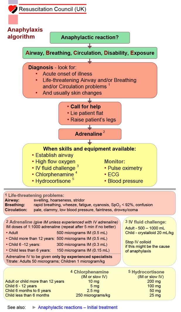
Anaphylaxis

Anaphylaxis


Adult Head Injury CT Guidelines - NICE

Adult Head Injury CT Guidelines - NICE


Children Head Injury CT Guidelines - NICE

Children Head Injury CT Guidelines - NICE


Adult C Spine CT Guidelines - NICE

Adult C Spine CT Guidelines - NICE


Traffic light system for identifying risk of serious illness in children - NICE

Traffic light system for identifying risk of serious illness in children - NICE


Kindly suggest if you have some more on your list, I will add in here.


Important Flow Charts

Thursday 30 July 2015

Major Incident Management

Major Incident Management is a very common topic these days for FCEM OSCE as well as SAQ papers. I have written here the strip down version and the basics which will be sufficient for the exam.


Classification of major incident:

• A simple major incident is one in which the infrastructure of the community in which it occurs remains intact, e.g. a train or air crash

• A compound major incident destroys or damages the infrastructure of the surrounding community

• A compensated major incident is one in which there are sufficient local resources to deal with the consequences

• An uncompensated major incident is one where the medical and other responding emergency services are destroyed or totally inadequate.


CSCATTT


The mnemonic describes a system widely accepted in the UK, and now in many other countries, which is designed to ensure the successful medical management of a major incident with live casualties. It is a hierarchy of actions that help the otherwise potentially chaotic actions of multiple staff to come together into a system.


The sequence can be remembered using the acronym "Command Spells Calm And Time To Treat". The overriding aim of this approach is to achieve the common aims of all emergency services at a major incident.


CCOMMAND


SSAFETY


CCOMMUNICATION


AASSESSMENT


TTRIAGE


TTREATMENT


TTRANSPORT


Responsibilities of the first crew on scene:


An ambulance is likely to be at the scene at an early stage. If it is a major incident, the crew should not get involved in treating individuals but they need to assess the situation and report back to control. There will be an Ambulance Incident Officer (AIO) who is the senior crew member who is in charge until a more senior officer arrives. His tasks include:


  • Assessing the scene.

  • Declaring a major incident and giving a situation report (SITREP or METHANE).

  • Deciding where to locate the Control Point, Casualty Clearing Station (CCS), and Ambulance Parking Point, as well as planning ambulance entry and exit routes.

  • The AIO is in charge of communication with all health service personnel on the scene.

  • The AIO discusses with the chain of command the need for additional support, such as a Medical Incident Officer (MIO) on scene, the Medical Emergency Response Incident Team (MERIT) and additional equipment.

Reporting a major incident


This must go through the appropriate channels so that all necessary personnel and services are informed. Full and relevant information must be gathered. There are two mnemonics to help with this. They are METHANE and CHALETS and the contents are similar:


ajor incident declared
E xact location
T ype of incident, eg explosion and fire in a tall building, release of gas in the underground system
H azards - present and potential
A ccess - routes that are safe to use
N umber, type, severity of casualties
E mergency services now present and those required.


C asualties - number, type, severity
H azards present
A ccess routes that are safe to use
L ocation
E mergency services present and required
T ype of incident, as above
S afety.


By now there may be several ambulances on the scene but the control vehicle is recognised as the one that still displays its flashing lights.


The medical emergency response incident team (MERIT)


This team (formerly known as the Mobile Medical Team) usually consists of a doctor and a nurse or two of each. They should stay together unless ordered to do otherwise. Ideally, they should not come from the hospital that will be receiving casualties as they need all their staff but, in a remote area, this may not be practical. They should arrive equipped with kit bags. These contain limited airway, breathing and circulation equipment. 


When the MERIT arrives at the scene they should report to the MIO whose position will be apparent from a flashing green beacon. If none is present, they should report to the AIO at the ambulance with the flashing blue light. The team will probably be sent to the CCS but may be required to assist with the triage and treatment of entrapped casualties. It is not the role of the doctor or nurse to get involved in search and rescue, counselling victims or commanding ambulance personnel.


Organisation at the scene and away


Overall control of the scene is the responsibility of the police who will control the outer cordon.


There will be a police manned incident control point through which all staff should enter and leave; all movements will be logged


If hazards are present, the Fire service will have responsibility inside the inner cordon (the hot zone) until the danger is controlled


Personnel entering and leaving the inner cordon must also be recorded for safety purposes


The bronze ( operational) area lies within the inner cordon and is the area where the rescue operation is in place. There will be bronze commanders ( forward commanders) from each emergency service. It is a dangerous area and medical activity within it is limited to:


  • Primary triage.

  • Evacuation of casualties.

  • Treatment of trapped casualties.

Silver ( tactical) command consists of the area within the outer cordon. The commanders from each service will be within this area, although they may move in and out of the bronze zones


Gold ( strategic) command is removed from the scene – usually in the police HQ or local authority buildings – and is the location where the chief officers from each emergency service meet.


A doctor may also be required to certify death. Casualties are evacuated to the CCS that will be close to the scene yet at a safe distance and linked to the ambulance loading point. The CCS is for secondary triage, initial stabilisation and preparation for transportation to hospital.


Triage


Triage is a system for sorting casualties into priority for treatment by subsequent teams. It enables limited resources to be deployed efficiently. Treating a less critically ill patient could deny life-saving interventions to others who may die as a result. A form of rapid assessment is required and the triage sieve is usually employed. An experienced operator can perform this in about 20 seconds, so that it is possible to triage many people in a short time. Priorities are numbered 1 to 3 in descending order of need and are colour-coded as follows:


  • P1: immediate priority. It is those who will die without immediate lifesaving intervention. Colour code red.

  • P2: intermediate priority. They will also need significant interventions but can wait a few hours. Colour code yellow.

  • P3: delayed priority. They will need medical treatment, but this can safely be delayed. Colour code green.

  • Dead is a fourth classification and is important to prevent the expenditure of limited resources on those who are beyond help. Colour code black.

Walking wounded are automatically classified as P3. This is related to the motor score on the Glasgow Coma Scale and predicts favourable outcome. Time can be saved in the bronze zone by asking all who can proceed to the CCS unaided to do so, and automatically classifying them all as P3. This is a useful technique if the area is hazardous and it is necessary to clear it and move on the injured as soon as possible.


Casualties need to be labelled and the cruciate triage card is useful. It has 4 arms, coloured red, yellow, green and black and the appropriate arm can be displayed. It also facilitates change of category if required. If nothing else is available, write on the person"s forehead. Dead bodies should be left where they are, partly to avoid unproductive use of resources and partly because this may be a scene of crime.


It is quite common for up to 50% of patients to be triaged into too high a category, competing for limited resources. It is common for children to be placed in too high a category and old people in too low a category.


How to deal with the number of casualties


On site


By the time that the CCS is set up there may be adequate resources. As a general rule, advanced life support should not be performed in the bronze zone, as it is very labour intensive and the chance of success is limited. More lives will be saved by attention to others. At the CCS there may be enough staff to enable this to be performed without neglecting others who would benefit from immediate attention.


ED - the casualties


The first patients to arrive at the hospital A&E department are usually the least severely injured. This is because they are the most mobile and the more severely injured may require stabilisation before moving. Hence, the arrival of a large number of people who are not severely injured may be the trigger for the declaration of an emergency. Valuable resources such as ambulances should not be used to transport those who can use other means including cars, taxis or getting on a bus. Those who arrive in A&E have probably been triaged twice already but they will still require a further triage. A different system is required in hospital from in "the field". There is no uniform system but a common classification is resuscitation, major and minor.


Managing the casualty numbers in hospital


The hospital major incident plan can be activated either on the request of the Ambulance Service, or autonomously by the hospital. When a disaster is declared it is necessary to try to gauge the scale of the problem and to make a rough estimate of the number and nature of injuries. 


  • It is common practice for one hospital to be the reception centre for injuries whilst another sends out staff to the scene.

  • The receiving hospital needs all its staff on site.

  • A centre of operations is set up there with clinical and managerial input.

  • The clinical director should be a senior doctor with authority, who is not directly involved in the care of the injured, as he cannot do both jobs simultaneously. Hence, he or she will almost certainly not be the ED consultant or probably any surgeon.

  • The command and control centre will probably not be in ED, as it will be extremely busy there.

ED - the non-casualties


There will be the usual flow of patients from unrelated events in A&E. Someone has to deal with them or send them elsewhere. This may be to another hospital or asking GPs to cope.


  • If the system is stretched, patients from unrelated sources need to be put through the same triage procedures as those from the major incident.

  • It is inappropriate to send away someone with a serious medical need just because he or she is not a victim of the major incident.

  • It is still useful to note who was from the incident to help with inquires and also reflection on the adequacy of the operation afterwards.

Other hospital departments


All staff (and not just those skilled in the management of trauma) should report for duty when a hospital declares a major incident.


  • Other patients will still need care when perhaps their usual carers have been called away.

  • Someone may need to take the decision to discharge patients to free beds for new arrivals.

  • Routine admissions must be halted.

  • The extent to which this will need to be done depends on the anticipated number of admissions.

  • There should be a designated area where staff can report to in order to be assigned to their duties.

  • Standards of care must be maintained.


Major Incident Management

Saturday 18 July 2015

Atrial Fibrillation

Atrial fibrillation (AF) is the most common cardiac arrhythmia. It is rarely a one-time event. Patients who develop AF once tend to be predisposed to developing it again & again. Current research indicates that this is primarily due to a change in the expression of ion channels that is caused by fibrillation (which may be mediated by changes in intracellular calcium caused by rapid pacing). This electrical remodelling creates an electrical “substrate” that is conducive to producing reentry. Some of the characteristics associated with AF are:


  • Symptoms: palpitations, dyspnea, fatigue, decreased exercise tolerance, chest pain


  • ECG: lack of P waves, “irregularly irregular” timing between QRS complexes & pulse pressure. The QRS complex & T waves are typically normal in duration & shape


  • Risk factors: hypertension, age, CHF, previous AF


  • Associated Pathology: atrial dilation, fibrosis, myocyte apoptosis


  • Complications: thromboembolism (due to vascular stasis in left atrium) & stroke


  • Common Origin & Mechanism: in patients with heart failure, an electrical focus around a left pulmonary vein combined with an abnormal atria; ablation of tissue around the pulmonary vein is one form of current therapy for patients with combined AF & heart failure (Khan et al., 2008).


There are two approaches to the treatment of AF:


  • Rate control, allowing AF to persist but controlling the ventricular rate by drugs affecting the AV node ERP (Effective Refractory Period), and

  • Rhythm control – with cardioversion to normal sinus rhythm & chronic treatment with antiarrhythmic drugs to prevent the reoccurrence of AF.

Arguments in favour of rate control include:


  1. Its easily achievable in most patients

  2. Avoidance of use of antiarrhythmic agents with less desirable side effects/toxicity

  3. Risk of stroke can be reduced by anticoagulant therapy

Arguments in favour of rhythm control include:


  1. Rhythm control reduces the odds of thromboembolism

  2. It was thought that patients who remain in AF have a worse outcome than those treated with drugs that maintain a sinus rhythm.

What does the evidence say?
In 2008, the results of two studies, one in North America, and one in Europe were published in the New England Journal of Medicine. In both studies, rhythm control provided no advantage over ventricular rate control with respect to survival (Fig 15A). On the basis of these results, rate control is currently considered an equally “safe” approach for the treatment of AF, and rhythm control (if it is used) can be abandoned early if it is not fully satisfactory (e.g. the patient cannot tolerate the side effects of the drugs used to suppress AF). Surgical procedures (e.g. Mini Maze procedure) involving catheter ablation of ectopic foci around the pulmonary veins may also be successful in patients with otherwise relatively normal hearts.


Figure 15. Panel A: Kaplan-Meier Estimates of Death from Cardiovascular Causes (Primary Outcome). Among 1376 patients with atrial fibrillation and congestive heart failure who were followed for a mean of 37 months, 182 patients (27%) in the rhythm-control group died from cardiovascular causes, as compared with 175 patients (25%) in the rate-control group (hazard ratio, 1.06; 95% confidence interval, 0.86 to 1.30). (From Roy et al, 2008). Panel B: Kaplan-Meier Estimates of the Percentage of Patients Remaining Free of Recurrence of Atrial Fibrillation in the two treatment groups (hazard ratio for recurrence among patients in the amiodarone group, 0.43 [95 percent confidence interval, 0.32 to 0.57]). Follow-up began 21 days after randomization (designated day 0). (From: Roy et al, 2000).


Currently amiodarone appears to be superior to other antiarrhythmics in preventing the reoccurrence of AFib (Figure 15B).


Initial conversion from AF to sinus rhythm can be achieved by either DC defibrillation or by an i.v. bolus of Amiodarone or Flecainide. Patients who are placed on rate control treatment are typically given maintenance therapy with either verapamil, diltiazem, a beta blocker or digoxin (digoxin use is more common if the patient has systolic HF).


NICE Recommendation (2014)


Perform manual pulse palpation to assess for the presence of an irregular pulse that may indicate underlying atrial fibrillation in people presenting with any of the following:


  • Breathlessness/dyspnoea

  • Palpitations

  • Syncope/dizziness

  • Chest discomfort

  • Stroke/transient ischaemic attack. [2006]


Perform an electrocardiogram (ECG) in all people, whether symptomatic or not, in whom atrial fibrillation is suspected because an irregular pulse has been detected. [2006]


Assessment of stroke and bleeding risks

Use the CHA2DS2-VASc stroke risk score to assess stroke risk in people with any of the following:


  • Symptomatic or asymptomatic paroxysmal, persistent or permanent atrial fibrillation

  • Atrial flutter

  • A continuing risk of arrhythmia recurrence after cardioversion back to sinus rhythm. [new 2014]

Use the HAS-BLED score to assess the risk of bleeding in people who are starting or have started anticoagulation.


Stroke prevention


  • Do not offer stroke prevention therapy to people aged under 65 years with atrial fibrillation and no risk factors other than their sex (that is, very low risk of stroke equating to a CHA2DS2-VASc score of 0 for men or 1 for women). [new 2014]

  • Consider anticoagulation for men with a CHA2DS2-VASc score of 1

  • Offer anticoagulation to people with a CHA2DS2-VASc score of 2 or above, taking bleeding risk into account.

  • Anticoagulation may be with apixaban, dabigatran etexilate, rivaroxaban or a vitamin K antagonist.


When to offer rate or rhythm control

Offer rate control as the first‑line strategy to people with atrial fibrillation, except in people:


  • Whose atrial fibrillation has a reversible cause

  • Who have heart failure thought to be primarily caused by atrial fibrillation

  • With new‑onset atrial fibrillation

  • With atrial flutter whose condition is considered suitable for an ablation strategy to restore sinus rhythm

  • For whom a rhythm control strategy would be more suitable based on clinical judgement.

Rate control


Offer either a standard beta‑blocker (that is, a beta‑blocker other than sotalol) or a rate‑limiting calcium‑channel blocker as initial monotherapy to people with atrial fibrillation who need drug treatment as part of a rate control strategy.


Consider digoxin monotherapy for people with non‑paroxysmal atrial fibrillation only if they are sedentary (do no or very little physical exercise).


If monotherapy does not control symptoms, and if continuing symptoms are thought to be due to poor ventricular rate control, consider combination therapy with any 2 of the following:


  • A beta‑blocker

  • Diltiazem

  • Digoxin.

Do not offer amiodarone for long‑term rate control.


Rhythm control


Consider pharmacological and/or electrical rhythm control for people with atrial fibrillation whose symptoms continue after heart rate has been controlled or for whom a rate‑control strategy has not been successful.


Cardioversion


For people having cardioversion for atrial fibrillation that has persisted for longer than 48 hours, offer electrical (rather than pharmacological) cardioversion.


Consider amiodarone therapy starting 4 weeks before and continuing for up to 12 months after electrical cardioversion to maintain sinus rhythm, and discuss the benefits and risks of amiodarone with the person.


For people with atrial fibrillation of greater than 48 hours" duration, in whom elective cardioversion is indicated:


Both transoesophageal echocardiography (TOE)‑guided cardioversion and conventional cardioversion should be considered equally effective.


Management for people presenting acutely with atrial fibrillation


Rate and rhythm control


  • Carry out emergency electrical cardioversion, without delaying to achieve anticoagulation, in people with life‑threatening haemodynamic instability caused by new‑onset atrial fibrillation.

  • In people with atrial fibrillation presenting acutely without life‑threatening haemodynamic instability, offer rate or rhythm control if the onset of the arrhythmia is less than 48 hours, and start rate control if it is more than 48 hours or is uncertain.

  • Consider either pharmacological or electrical cardioversion depending on clinical circumstances and resources in people with new‑onset atrial fibrillation who will be treated with a rhythm control strategy.

  • If pharmacological cardioversion has been agreed on clinical and resource grounds for new‑onset atrial fibrillation, offer:

  • Flecainide or amiodarone if there is no evidence of structural or ischaemic heart disease or

  • Amiodarone if there is evidence of structural heart disease.

  • In people with atrial fibrillation in whom the duration of the arrhythmia is greater than 48 hours or uncertain and considered for long‑term rhythm control, delay cardioversion until they have been maintained on therapeutic anticoagulation for a minimum of 3 weeks. During this period offer rate control as appropriate.

  • Do not offer magnesium or a calcium‑channel blocker for pharmacological cardioversion.

Anticoagulation


In people with new‑onset atrial fibrillation who are receiving no, or subtherapeutic, anticoagulation therapy:


  • In the absence of contraindications, offer heparin at initial presentation

  • Continue heparin until a full assessment has been made and appropriate antithrombotic therapy has been started

Useful Videos

Useful Videos


Atrial Fibrillation

Sunday 3 May 2015

Emergency Department Clinical Quality Indicators

The Department of Health introduced five new headline Emergency Department (ED) clinical quality indicators from the 1 April 2011 with three supporting measures.


The five headline measures are:


  • Unplanned re-attendance rate – Patients who return to A&E within seven days of the original attendance are classed as an unplanned reattendance if they have not been specifically asked to re-attend.

  • Total time in the  accident and emergency department- This is measured from the time of arrival and registration on the hospital information system to the time that the patient leaves the department to return home or to be admitted to a ward bed.

  • Left without being seen rate- Patients who have registered but leave the department without waiting to be seen by a clinician.

  • Time to initial assessment – This applies only to patients who are brought in by ambulance and is measured from the time of arrival in the department to the time the patient has an assessment by the clinical team.

  • Time to treatment – This is the time from arrival for all patients to seeing a doctor or nurse practitioner who will start the treatment for the patient’s condition.

The three supporting indicators are:


  • Ambulatory care – Emergency departments are developing pathways of care for patients to avoid hospital admission and the two key areas of work at present are for patients who have Cellulitis and need antibiotics and for those who have DVT. The measure will reflect the proportion of patients who are able to be treated at home by these improved pathways and processes.

  • Service experience – One of the key measures of the service quality is the experience of the patient. Trusts will be reporting the results of patient surveys as well as giving information about specific projects designed to provide a better experience or outcome for the patients overall.

  • Consultant sign-off – While it is not possible or necessary for every patient to be seen by a consultant in A&E, there are certain conditions where the outcome can be improved by a senior doctor being involved in the case. The measure will demonstrate the proportion of those patients who have seen a senior doctor and will be measured twice a year by detailed review of notes and outcomes.


The patient groups for consultant sign-off:


  • Adults (over 17 years of age) with non-traumatic chest pain

  • Febrile children less than 1 year old

  • Patients making an unscheduled return to the ED with the same condition within 72 hours of discharge from the ED


Emergency Department Clinical Quality Indicators

Functions, Role and Regulations of Care Quality Commission (CQC)


CQC is England’s health and social care services regulator. They make sure health and social care services provide people with safe, effective, compassionate, high-quality care and we encourage them to improve.


CQC’s principles are to:


  • Put people who use services at the heart of its work

  • Have an open and accessible culture

  • Be independent, rigorous, fair and consistent

  • Work in partnership across the health and social care system

  • Commit to being a high-performing organisation

  • Promote equality, diversity and human rights

What does the CQC do?


CQC carries out its role in the following ways:


  • Setting national standards of quality and safety that people can expect whenever they receive care.

  • Registering care services that meet national standards.

  • Monitoring, inspecting and regulating care services to make sure they continue to meet the standards.

  • Protecting the rights of vulnerable people, including those whose rights are restricted under the Mental Health Act.

  • Listening to and acting on patient concerns and experiences.

  • Working in partnership with other organisations and local groups.

  • Challenging all providers, with the worst performers getting the most attention.

  • Making fair and authoritative judgements supported by the best information and evidence.

  • Taking appropriate action if care services are failing to meet the standards.

  • Carrying out in-depth investigations to look at care across the system.

  • Reporting on the quality of care services, publishing clear and comprehensive information, including performance ratings to help people choose care.

Who do CQC regulate?


CQC regulate:


  • Treatment, care and support provided by hospitals, GPs dentists, ambulances and mental health services.

  • Treatment, care and support services for adults in care homes and in people’s own homes (both personal and nursing care).

  • Services for people whose rights are restricted under the Mental Health Act.

How are bodies regulated?


In accordance with ‘The Scope of Regulation – August 2013’ CQC Guidance Leaflet, the CQC say that all health and social care services in England need to register with them. In practice however, this is confined to those who carry out any of the CQC’s fifteen regulated activities.


These regulated activities are listed in Schedule 1 of the Health and Social Care Act 2008 (Regulated Activities) Regulations 2012. These include:


  1. Personal care (which is provided to them in the place where those people are living at the time when the care is provided)

  2. Accommodation for person who require nursing or personal care

  3. Accommodation for persons who require treatment for substance misuse

  4. Accommodation and nursing or personal care in the further education sector

  5. Treatment of disease, disorder or injury (if provided by a listed health care professional, social worker or multi-disciplinary team)

  6. Assessment or medical treatment for persons detained under the Mental Health Act 1983 (only in hospitals)

  7. Surgical procedures (carried out by a healthcare professional, some exceptions for minor procedures, eg foot and nail treatment, tattooing, piercing, removal of hair and blemishes, etc)

  8. Diagnostic and screening procedures (radiation, ultrasound and MRI etc)

  9. Management of supply of blood and blood-derived products (includes NHS Blood and Transplant)

  10. Services in slimming clinics (that include giving medicines AND are led by a registered medical practitioner)

  11. Transport services (where the vehicles primary purpose is for transporting people who require treatment), triage and medical advice provided remotely (where the advice is medical AND responsive i.e. for immediate attention or action OR it constitutes triage AND is provided by a body responsible for that service)

  12. Maternity and midwifery services

  13. Termination of pregnancies (does not include the ‘morning after pill’ when used as an emergency contraceptive)

  14. Nursing care (where not under another regulated activity)

  15. Family planning services (services for the insertion or removal of an intrauterine contraceptive device by, or under the supervision of, a health care professional)

Any organisation that carries out these regulated activities in England must register with CQC. Separate provisions are available in the rest of the UK.


For any bodies that carry out these services in England from neighbouring countries for short periods of time (eg. Ambulance services) the CQC have said that they will take a ‘proportionate’ and ‘reasonable’ approach to registration.


Registration takes many forms and varies slightly for different practice groups (eg. Corporate groups, franchises, partnerships). This includes the fact that a GP surgery is normally registered under their partner’s (lead doctor’s) name, rather than the surgery itself.


Failure to register


Any service that provides one or any of the fifteen regulated activities who fails to register will be liable for a criminal offence and may face prosecution.


Section 10 of the Health and Social Care Act 2008 states:


(1) Any person who carries on a regulated activity without being registered under this Chapter in respect of the carrying on of that activity is guilty of an offence.

(4) A person guilty of an offence under this section is liable—


  • on summary conviction, to a fine not exceeding £50,000, or to imprisonment for a term not exceeding 12 months, or to both;

  • on conviction on indictment, to a fine, or to imprisonment for a term not exceeding 12 months, or to both.

Exemptions to regulation


The following bodies aren’t covered currently, even though they provide regulated activities:


  • Scottish, Irish, Welsh and non-Mainland UK services

  • Non-NHS primary pharmaceutical services – eg. Boots, Superdrug, Lloyds

  • Non-NHS primary ophthalmic services – eg. Specsavers, Vision Express

  • Some mobile treatment units – eg. Blood donation, breast scanning. Often broader registration is kept with the CQC.

  • Some fertility clinics after 01 October 2013

  • Independent practitioners – acting as a ‘designated body’ providing services in a surgery or consulting room, treatment does not include anaesthesia or intravenous sedation, childbirth services and termination of pregnancy, certain cosmetic surgery, haemodialysis etc.

  • Independent midwives - exemption if midwife is self-employed AND providing non-NHS care AND providing services to their patient ONLY in their home.

  • Third party exemptions - Occupational health schemes; defence medical/dental teams for the armed services; forensic medical service (eg in police custody); medical assessment or treatment linked to insurance schemes (does not exclude services through private medical insurance schemes); medical services organised by a government department that do not involve treatment requiring admission to hospital (eg. medical assessments to determine eligibility for social security benefits).

  • Individual budgets, individual user trusts and self-funded personal care or nursing care - Where a person, or a related third party on their behalf, makes their own arrangement for nursing care or personal care, and the nurse or carer works directly for them and under their control without an agency or employer involved in managing or directing the care provided, the nurse or carer does not need to register for that regulated activity.

  • Any health or social care activity carried out by a carer for a member of their family or someone in a personal relationship – where there is no commercial consideration.

  • School nurses - who are employed and managed by the school and who provide services to the school"s pupils. In general, this will exempt school nurses in independent schools, but not in public sector schools where the school nursing service will be included in the registration of the relevant provider.

  • Aircraft operators – that only carry out the activity of transport services and do not carry out other regulated activities (such as treatment of disease, disorder or injury)

  • Aircraft operators or air ambulance providers within the confines of event grounds - may fall out of regulation if their transport is confined to the event ground.

Archived services


Beyond these exempted services, the CQC have also removed a number of healthcare providers from their website.


These services include those where:


  • The provider has voluntarily closed it – for example, a service closed because it is no longer profitable.

  • The CQC has taken enforcement action to close it – for example, a service whose registration is cancelled after CQC inspectors found evidence of poor care or abuse.

  • The legal entity providing the service has changed – for example, a service where a sole trader has been replaced by a partnership as the provider.

  • The service has changed address - for example, a service that has moved to a new building to take advantage of better facilities.

  • The provider is no longer required to be registered with the CQC - for example, some fertility clinics that have no longer been regulated by the CQC since 01 October 2013.

This means that it is very important to record the correct name of the healthcare provider when taking feedback from a helpline caller. Common mistakes may include recording a trust under a generic name (eg ‘Manchester Hospital’) or failing to specify which trust is being discussed.


What are the potential risks for service users?


When services are exempted by CQC, service users cannot feedback their experiences. This means that if poor care is experienced, or members of the public know that poor care is being provided, the information cannot be collected and acted upon by the CQC, these cases are not included in the CQC pilot.


CQC continues to expand in the services it regulates and covers. Changes to the CQC regulations were made following a consultation by the Department of Health in 2011. These amendments were then approved by Parliament in 2012. These changes mostly address technical points in the regulations that make them clearer in order to provide greater clarity.


Further changes in October 2013 meant that some further healthcare providers were no longer required to be registered with the CQC. The CQC have responded by saying that many of these services were already exempted by the regulations by another means.


With input from AVMA


Functions, Role and Regulations of Care Quality Commission (CQC)